
Seeing Better, Knowing More: Advanced Imaging Enables Diagnosis and Treatment
05.12.2010
Eleanor Walker, 88, has played golf for more than three decades. It's the kind of exercise that has kept her energy at a level far beyond her years.
Eleanor Walker has played golf since she was in her 50s. She saw no reason to stop when she turned 88 this year. Sure, she took a bit more time getting her clubs out of her car, and she conceded that a golf cart should be her transportation around the course. Other than that, she was just fine.
This spring, before the weather was reliable enough for golf, she had lunch at the course clubhouse with her sister-in-law. After lunch, the two women came out to their car. The next thing Walker remembers is being in an ambulance. At least, she thought she was in an ambulance. Things were a bit confused. Then, she remembers feeling pretty relaxed and people were asking her questions, asking her to move this and that.
Now she knows she had had a stroke—that within her brain, a clot had blocked blood flow to one of her arteries and its many branches. Parts of her brain were no longer getting the oxygen they needed to function. When she arrived at Stanford Hospital, she could not speak or move her right side. If the clot was not removed, the damage would continue, claiming more and more of her brain and leaving her with less and less ability to be the fully active woman she had been.
What she didn't understand at the time was the unique combination of imaging and expertise that was ready and waiting for her at Stanford Hospital & Clinics. Or how, precisely, that combination's powerful synergy gave her back her life, just as it had been, in spite of odds that were not particularly in her favor.
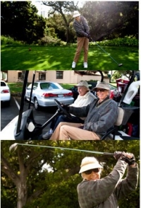
After prompt treatment at Stanford Hospital's nationally recognized Stroke Center, Eleanor Walker, 88, is back to her regular golfing routine, which includes driving herself from her home to the course and playing a full round, sometimes with a friend.
Faster, Smaller, Clearer
If it had been 1980, Walker's outcome might have been very different. Her physicians would have seen Walker's brain with images that recorded a miniscule fraction of what they can see now. And they would have waited for those images thousands of times longer than they do today. In the past 30 years, medical imaging's increase in speed and data has matched that of computers. Instead of one image recorded about every four seconds, today's imaging technology captures 320 images in 0.3 seconds. Like a jet versus an ant. And instead of seeing just a beach in that jet's flyover, today's imaging technology sees grains of sand.
Roughly 75 percent of our brain is wired for vision, so images are a powerful tool.
At Stanford's Department of Radiology, where clinical diagnosis and treatment reflect the latest research data, physicians are using the most advanced methods of imaging to push breakthroughs in several areas, working in collaboration with engineers and physicists. Once X-rays were the best medicine could offer. Now, physicians use ultrasound, computed tomography, magnetic resonance, optical bioluminescence and fluorescence to bear down on body tissue even to its molecules. Recently, Stanford radiologist Sanjiv Sam Gambhir, MD, PhD, pioneered the use of another form of molecular imaging, one to track, even more accurately, the biochemical changes that mark cancer before its structural changes are visible.
These advances have affected every aspect of medicine, saving many, many lives. Patients can be diagnosed earlier, vastly improving the chance of recovery. Physicians can plan more accurate treatment, without the kind of exploratory surgery that was once the only way to see what was going on. They can work inside the body in ways previously not dreamed possible.
At Stanford, Walker's physicians quickly scanned her brain to find the clot. Within a couple of minutes, 500 to 1,000 images in that scan were fed through Stanford's specially-developed software to create special images that showed how much blood was flowing through the arteries to her brain and how long it took to get there. That information answered the most important questions about Walker's condition. How much of her brain had the stroke already damaged−and how much might soon be? Could rapid treatment give Walker a good chance for a nearly complete recovery? And could it be done safely?
Seeing Every Step
The ability of physicians to see inside the body, with the kind of detail imaging technology now supplies, has allowed a whole new set of non-surgical, minimally-invasive treatments and an expanded role for radiologists. Among Walker's team of physicians were neurologists, diagnostic radiologists, and an interventional radiologist, Michael Marks, MD, who took the next clinical step in treating Walker. Interventional radiologists, like Marks, enter the body guided by imaging technology, using small tubing called catheters, not scalpels, eliminating traditional surgery's risks from large incisions.
I asked my doctor if I could play golf and she said, 'Sure you can!' Maybe I'll even play better!
To get to Walker's brain, Marks made a tiny incision in an artery at the top of her leg and into that incision threaded a tiny catheter, inch by inch, following the artery into the brain, all the while guiding its progress by watching an image of that artery captured on video. The catheter carried a tiny, corkscrew-like wire inside its narrow tubing. Once Marks saw the catheter was at the clot, he turned the wire to grab hold of the clot and removed it. On the video, he could see the blood again fully flowing through the artery.
A couple of days after Walker's stroke, she was back home. A couple weeks later, she was out playing golf again. "I asked my doctor if I could play—and she said, 'Sure you can!'" Walker said. "I used to be able to do everything so fast. I'm a little slower now. I'll just have to practice. Maybe I'll play even better!"
More and more, almost every step of medical care is enhanced by imaging. Stanford cancer physicians use advanced imaging technology to plot and then deliver precisely targeted radiation in robot-assisted procedures. In order to treat lung cancer, difficult because tumors move with each breath a patient makes, the CyberKnife follows imaging information from real-time video. Imaging also enables the CyberKnife and other high-beam radiation devices to treat the brain without damaging surrounding tissues.
And when surgeons choose to treat patients with small-incision or minimally invasive surgery, optics in the instruments they use are essential, and improving all the time.
Walker's brain information came from an advanced CT tailored for acute stroke patients at the Stanford Stroke Center, one of the first in the U.S. to offer stroke prevention, treatment and research. The Stanford protocol has made a real difference for people like Walker, whose age and functional effect from the stroke might have eliminated her as a candidate for aggressive care without the special information available at Stanford. The analytical software to construct an image of the brain tells physicians and families much more quickly what they need to know about what's possible for a stroke patient. "There are few places in the U.S. that use it in the acute way we do," said radiologist Greg Zaharchuk, MD. "That software means you can get so much more information from that CT."
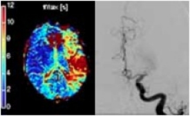
When Eleanor Walker arrived at Stanford Hospital, physicians immediately looked at her brain with a CT scanner to find the clot and to see what damage had already been done. They also put contrast dye into her artery and recorded images of its path with X-ray in a process called angiography. Digital subtraction removes everything in the image except the artery marked with the contrast material. These images were made before her treatment.
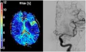
What a clot does to blood flow is easy to see in these images, taken after interventional radiologists removed the clot and restored normal passage of blood flow to Walker's brain. The images on the left mark, with colors, how long blood is taking to get to various parts of the brain. The red areas mark where blood flow has stopped, the blue where blood flow is normal, the yellow and green where damage will soon occur.
* In addition to its imaging availibility at the Hospital, Stanford's Department of Radiology has two other centers - at the Outpatient Center in Redwood City and the Stanford Medicine Imaging facility in Palo Alto.
Expanding View
Accomplishing the image goal with a CT, instead of an MRI, means patients can be assessed more quickly. The software also automates the information analysis, a big improvement over the previous state where someone had to be called to physically come to the Hospital to run a program.
The treatment of other brain conditions, like epilepsy and Parkinson's, is also becoming more effective with imaging and software analysis Stanford physicians have developed to understand the brain's behavior in greater detail. That imaging-assisted mapping also guides cardiovascular surgeons as they repair or replace heart valves. The special software they use, which can build 3D images to track blood flow and volume, gives an instant answer about the effectiveness of that repair or replacement.
Soon, imaging could make it possible to see the cerebral plaque that identifies the presence of Alzheimer's. Physicians at Stanford already use imaging techniques that help them place radiation-loaded microspheres directly into veins feeding tumors in the liver, a direct approach that avoids the drastic effect of chemotherapy delivered throughout the body.
As might be expected, scanning the brain to create images of what it looks like when people think, feel or do certain things, is proving to be a cornucopia of information. Recently, brain scans revealed more about what goes on in anxiety disorders and post-traumatic stress disorder. Understanding the relationships between the various parts of the brain takes physicians one step closer to therapeutic solutions.
The collaboration of science, physics and engineering, said Gary Glazer, MD, Chair of Stanford's Department of Radiology, is especially strong at Stanford. And that kind of collaboration drives the future of imaging. At the heart of it, though, is the very nature of human physiology. "What we do is try to explain complex systems through visual representation," he said. "Roughly 75 percent of our brain is wired for vision, so images are a powerful tool to understand that complexity."
Walker is getting stronger each day, back to her old self. "I don't really pay attention to my age," she said. "I just put it out of mind." Her memory of what happened will likely always be spotty. What she does recall best is how relaxed she felt in the Hospital. "I just didn't worry about it. I assumed I would be fine."
CARE AT STANFORD
As the world’s first designated Comprehensive Certified Stroke Center, we offer the highest level of stroke prevention and care for all causes of stroke.
Visit Stroke Center »