Learn about the flu shot, COVID-19 vaccine, and our masking policy »

New to MyHealth?
Manage Your Care From Anywhere.
Access your health information from any device with MyHealth. You can message your clinic, view lab results, schedule an appointment, and pay your bill.
ALREADY HAVE AN ACCESS CODE?
DON'T HAVE AN ACCESS CODE?
NEED MORE DETAILS?
MyHealth for Mobile
Get the iPhone MyHealth app »
Get the Android MyHealth app »
How does a Targeted Biopsy Work
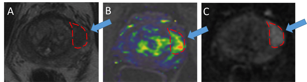
Multiparametric MRI—Representative case demonstrating the value of targeted prostate biopsy in a 60 year old man with a high blood level of PSA. He underwent three conventional prostate biopsies over 5 years that did not detect prostate cancer. During this time his PSA continued to rise. At Stanford, a multiparametric prostate MRI showed an area that was highly suspicious for cancer on T2-weighted imaging (A), contrast-enhanced imaging (B), and diffusion weighted imaging (C). Based on this result, a targeted prostate biopsy was scheduled.
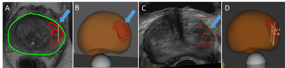
Targeted prostate biopsy—During the biopsy, the MRI (A) was fused with ultrasound to generate a 3-dimensional model (B) of the prostate that included the abnormal area from the MRI. By fusing MRI with ultrasound (C), the urologist targeted several biopsy needles directly into the suspicious area (D).