Meningioma
A meningioma is a slow-growing tumor that develops in the covering of the brain or spinal cord (meninges). Meningiomas are usually not cancerous and may not require treatment unless they press on surrounding brain tissue, nerves, or blood vessels.
The Stanford Health Care Brain and Spine Tumor Program is known for innovation, collaborative multi-specialty care, and compassion. It is a global referral center for the diagnosis and treatment of meningiomas.
What is a meningioma tumor?
A meningioma is a tumor that forms in the meninges, the three layers of tissue that cover the brain and spinal cord. Meningiomas are the most common type of brain and spine tumor.
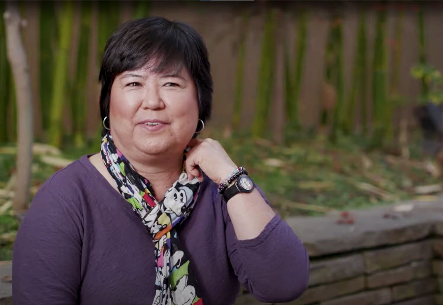
Most meningiomas are benign (noncancerous), and small tumors that don’t cause symptoms may not require treatment. If the meningioma worsens and needs treatment, our doctors use two main approaches: surgery and radiation therapy. Learn more about meningiomas in this video.
Connect to Care
Let us help find personalized care options for you and your family.
Understanding Meningiomas
Meningioma Symptoms
People with a meningioma brain or spine tumor often experience no symptoms at all. Doctors sometimes discover the tumor while examining the brain or spine for another health problem.
Symptoms from a meningioma vary depending on the tumor’s size and location. Meningioma symptoms may include:
- Blurred vision, double vision, or loss of peripheral (side) vision
- Difficulty with balance
- Headache
- Memory or hearing loss
- Seizures
- Weakness or numbness in arms or legs
Types of Meningioma
Doctors classify meningioma tumors according to where they form. Most meningiomas are around the brain, but some develop in the meninges covering the spinal cord.
Types of meningiomas that form near the skull base include:
- Posterior fossa/petrous meningiomas form underneath the brain and press on the trigeminal nerve, triggering a condition called trigeminal neuralgia.
- Suprasellar meningiomas start in the skull base near the pituitary gland and optic nerve, often causing vision and pituitary problems.
- Skull-base meningiomas form in the bones in the bottom of the skull or in the boney ridge behind the eyes.
- Sphenoid wing meningiomas form in the skull base behind the eyes.
Types of meningiomas that develop elsewhere in the brain include:
- Convexity meningiomas grow on the brain’s surface.
- Falcine and parasgittal meningiomas form in or near the thin layer of tissue between the two sides (hemispheres) of the brain.
- Intraventricular meningiomas form in the part of the brain that produces and distributes spinal fluid.
- Olfactory groove meningiomas form along the nerves that connect the brain and the nose and may affect smell and vision.
- Recurrent meningiomas are meningiomas that return after treatment. They may share the same characteristics as the first meningioma or act more aggressively.
Meningioma Risk Factors
In most cases, we don't know what causes meningiomas. However, some factors may increase your risk, including:
- Age: Meningioma is most common in people age 65 and older.
- Gender: Women are more likely to develop noncancerous meningioma than men. Cancerous meningiomas occur equally in men and women.
- Radiation: Head exposure to radiation, such as radiation therapy, may increase risk.
- Family history: An inherited (passed down in families) genetic condition called neurofibromatosis type 2 may increase the risk for meningiomas, particularly cancerous tumors.
Grades of Meningioma
Although doctors typically categorize brain tumors into four grades, meningiomas are classified into only three. Doctors determine the grade based on the cells’ appearance under a microscope.
Meningioma grades include:
- Grade 1 meningioma (benign): Cells look the least abnormal. Most benign meningiomas are successfully treated with surgery.
- Grade 2 meningioma (invasive or atypical): Cells look a bit more abnormal. These tumors are more likely to grow and spread into surrounding tissue.
- Grade 3 meningioma (anaplastic): Cells look the most abnormal. Such tumors typically grow quickly and are the most likely to return after treatment.
Diagnostic Tests for Meningioma
At Stanford, we tailor the diagnostic phase of brain tumor care to each person. If we need further testing to confirm a diagnosis, your doctor and care team will work with you to determine which tests you need. Tests may include:
Medical and Family History
To diagnose meningioma, your doctor typically starts by asking about your medical history, including any previous radiation therapy, and lifestyle habits. Sharing your family’s medical history also helps doctors understand your situation and alert them to possible inherited genetic disorders.
Neurological Examination
During a neurological exam, your doctor looks for changes in your vision, hearing, balance, coordination, strength, and reflexes. These changes may help determine the tumor’s location.
If you have had screening elsewhere and received abnormal results, we may perform additional imaging, if needed. Brain scans provide detailed images that give us the most precise understanding of the tumor.
Your doctor may perform a biopsy to confirm a meningioma diagnosis. A biopsy also provides information to help guide treatment decisions.
During a biopsy, your doctor uses a small needle to remove cells from the tumor. Doctors look at the cells under a microscope to see if the tumor is cancerous. They also determine its grade, indicating how aggressive it is.
Leaders in Skull Base Surgery
Skull base surgery is a highly-specialized field that addresses tumors and other abnormalities on the underside of the brain or base of the skull. Our program is ranked among the top in the world.
Meningioma
Meningiomas are slow-growing tumors in the tissues around the brain. Learn about meningioma symptoms and our advanced methods for diagnosis and treatment.
meningioma
meningioma tumor
meningioma brain tumor


