
PET/CT Scan
Stanford is the first health care institution in Northern California to offer patients a powerful new diagnostic imaging system known as Positron Emission Tomography/Computerized Tomography (PET/CT) scanning.
This hybrid technology combines the strengths of two well-established imaging modalities in one imaging session to more accurately diagnose and locate cancers while increasing patient comfort.
Today, most PET scans are performed on instruments that are combined PET and CT scanners. The combined PET/CT scans provide images that pinpoint the location of abnormal metabolic activity within the body, like malignant tumor cells. The combined scans have been shown to provide more accurate diagnoses than the two scans performed separately.
Every PET/CT scan at Stanford is reviewed and correlated by both a board certified nuclear medicine doctor and a board certified radiologist at a daily joint review session. Separate full reports are generated from each division for each patient.
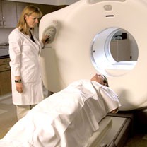
The PET and CT scans are done at the same time on the same machine. The physician is able to precisely overlay the metabolic data of the PET scan and the detailed anatomic data of the CT scan to make a more detailed image than either test would make by itself. A small amount of radioactive glucose (sugar) is injected into a vein. The PET scanner rotates around the body and makes a picture of where glucose is being used in the body. Malignant tumor cells show up brighter in the picture because they are more active and take up more glucose than normal cells do. Some people are sensitive to the radioactive glucose and may have nausea, headache, or vomiting.
PET, or positron emission tomography, monitors the biochemical functioning of cells by detecting how they process certain compounds, such as glucose (sugar). Cancer cells metabolize glucose at a much higher level than normal tissues.
By detecting increased glucose use with a high degree of sensitivity, PET identifies cancerous cells—even at an early stage when other modalities may miss them. However, PET cannot pinpoint the exact size and location of tumors to a precision necessary for optimal diagnosis and treatment planning.
Learn more about PET scans.
CT, or computed tomography, yields a detailed picture of the body's anatomical structures by taking cross-sectional images or X-ray slices of the body. While CT does an excellent job of depicting structures and anatomy, it may miss small or early stage tumors.
Learn more about CT scans.
Currently, doctors can overlay the results of PET and CT scans performed separately to identify and locate tumors. However, because a patient may not be positioned identically for both scans, the two images can be difficult to line up exactly, degrading the accuracy of the diagnostic information.
The combined PET/CT machine allows doctors to rapidly perform both scans in one session without having to move the patient. This means doctors can precisely overlay the metabolic data of the PET scan and the detailed anatomic data of the CT scan to pinpoint the location and stage of tumors.