
Lung Volume Reduction Surgery for Emphysema (LVRS)
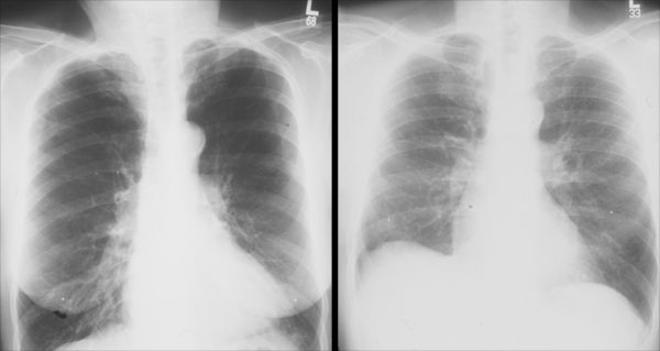
Chest X-rays before and after LVRS. Note the flattened diaphragms at the bottom of the chest and dark, “radiolucent” lungs before the operation, both of which are improved on the postoperative chest x-ray.

Intraoperative photo showing the stapling device used to remove the most abnormal, emphysematous lung tissue. Note the white “buttress” material used to prevent air leaks from the lung.

Typical areas of lung removed during LVRS
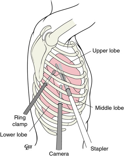
Incision placement for the thoracoscopic (VATS) approach to the operation
Read more about lung volume reduction surgery for emphysema and view Dr. Shrager's video.
Other Clinics
Lung Volume Reduction Surgery
Lung volume reduction surgery is a treatment for emphysema, a lung disease (often caused by smoking) which prevents oxygen from reaching the bloodstream.
lung volume reduction surgery
emphysema treatment
LVRS