The Role of White-Light 3D Scanning in Treatment of Clavicle Fracture
12.23.2020
Stanford Medicine orthopaedic surgeons have identified a new way to evaluate and monitor shoulder deformity after displaced diaphyseal clavicle fracture (DCF). White-light 3D body scanning allows surgeons to quantify clinically relevant shoulder girdle deformity with more accuracy than radiographic assessment or manual measurement. This information may further guide clinical decisions about operative versus nonoperative management of DCF.
Importance of 3D body scanning for displaced fractures
According to Michael Gardner, MD, chief of the orthopaedic trauma service, radiographic parameters historically informed clinical decision making for DCF. Yet, measurements taken this way fail to capture complex 3D changes in the shoulder associated with a clavicle fracture. While CT offers a more accurate assessment of deformity, it is not routinely performed due to radiation exposure and cost.
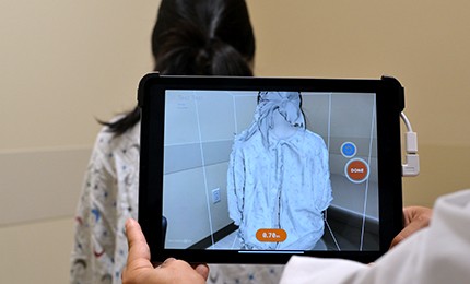
White-light scanning technology provides a detailed 3D representation of the anatomy, allowing surgeons to accurately measure the differences between injured and uninjured shoulders. Accurate evaluation is key to avoiding associated multiplanar deformity above the shoulder girdle that often follows nonsurgical treatment of DCF.
Dr. Gardner suggests the small 3D body scanner can potentially be used anywhere in the hospital or even at the scene of the trauma to deliver findings quickly. Additional benefits of digital scanning for shoulder deformity include:
- No exposure to radiation
- Less costly than radiographic or CT imaging
Evaluation 3D scanning for clavicle fractures
Aided by a grant from the Orthopaedic Trauma Association, Dr. Gardner and other Stanford Medicine researchers studied the efficacy of white-light 3D body (digital) scanning in 22 adult patients with DCF (OTA/AO 15A). After identifying specific anatomic landmarks to be measured, investigators used artificial intelligence (AI) to create measurements for both injured and uninjured shoulders.
When compared with manual measurements and radiographic imaging, digital scanning showed high sensitivity in identifying shoulder deformity, according to Dr. Gardner. White-light 3D scanning detected significant, clinically relevant differences between injured and uninjured shoulders. Neither the obtained manual measurements nor radiographic imaging accurately recognized those deformities.
For patients with DCF, physicians may use this additional data in ways that include:
- Informing treatment decisions (surgical versus nonsurgical management)
- Monitoring anatomical restoration (in surgical patients)
- Monitoring ongoing deformity (in nonsurgical patients)
The role of digital scanning in orthopaedic surgery
While 3D scanning is a useful tool in determining shoulder deformity, it is considered supplemental to clinical examination and radiographic assessment. As a new technology, 3D scanning has not been thoroughly evaluated as a diagnostic imaging tool for musculoskeletal trauma.
Outside of clavicle fractures, Dr. Gardner expects white-light 3D body scanning may eventually have a role in other orthopaedic capacities, including scoliosis evaluation and limb measurement.
AI technology in orthopaedic trauma
Identifying shoulder deformity following DCF is not the only application for AI within orthopaedic trauma at Stanford Medicine. Researchers are currently investigating how to use complex algorithms and AI to predict better outcomes following hip and femur fractures.
Through AI technology, orthopaedic surgeons like Dr. Gardner hope to accurately identify patients at higher risk of complication or mortality during the preoperative period. Stanford Medicine researchers conduct retrospective studies to understand surgical outcomes. AI technology provides risk stratification using the data collected, which may inform treatment decisions and considerations.
Referring patients to Stanford Health Care
Learn more about referring patients to the Orthopaedic Trauma Service at Stanford Health Care.
Find out more about our current clinical trials.
Clinical Trials

Learn more about the latest advanced clinical trials in Orthopaedics and Sports Medicine at Stanford Health Care and discover open trials currently accepting patients.