
What Is Tomosynthesis (3D mammography)?
Tomosynthesis or “3D” mammography is a new type of digital x-ray mammogram which creates 2D and 3D-like pictures of the breasts. This tool improves the ability of mammography to detect early breast cancers, and decreases the number of women “called back” for additional tests for findings that are not cancers.
During a “3D” exam, an X-ray arm sweeps in a slight arc over your breast, taking multiple low dose x-ray images. Then, a computer produces synthetic 2D and “3D” images of your breast tissue. The images include thin one millimeter slices, enabling the radiologist to scroll through images of the entire breast like flipping through pages of a book, and providing more detail than previously possible.
The “3D” images reduce the overlap of breast tissue, and make it possible for a radiologist to better see through your breast tissue on the mammogram.

With conventional digital mammography, the radiologist is viewing the tissues of your breast overlapping on flat images. This tissue overlap can sometimes make cancers hard to detect. Also, overlap can sometimes create areas that appear abnormal, but require that you be “called back” for additional tests to determine that cancer is not present (so-called false positives).
Tomosynthesis or “3D” mammography directly addresses the current limitations of standard 2D mammography. Multiple studies have shown that “3D” mammography increases the detection of breast cancer by approximately 25%, and decreases the number of false positive call backs by approximately 15%.
A screening mammogram is done in women who have no breast signs symptoms. A diagnostic mammogram is done in women who have been “called back” from a screening mammogram, or who have a clinical breast symptom such as a lump.
Having a “3D” mammogram is similar to a having conventional digital mammogram, including the amount of compression of the breasts and the time in compression. The main difference is that the X-ray arm sweeps in a slight arc over your breasts.
- Decreases radiation dose
- Separates glandular tissue
- Decreases superimposition of tissue
- Improves resolution or clarity of the image
- Increases contrast to visualize subtle differences in tissue
- Reduces scatter radiation
It is approved for all women who would be undergoing a standard mammogram, in both the screening and diagnostic settings.
Because Stanford has invested in software that creates both the synthetic 2D and “3D” images from the same acquisition, the synthetic 2D and “3D” radiation dose is very similar to that of standard 2D digital mammograms in the USA.
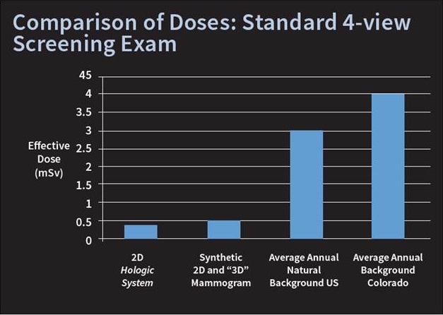
- The average annual natural background in the U.S. is 3 millisieverts (mSv). In Colorado it is 4 mSv.
- A traditional 2D mammogram is 0.4 mSv.
- A synthetic 2D and “3D” mammogram is 0.5 mSv.
Sources:
1 Bernardi D, Ciatto S, Pellegrini M, et. al. Prospective study of breast tomosynthesis as a triage to assessment in screening. Breast Cancer Res Treat. 2012 Jan 22 [Epub ahead of print].
2 The Hologic Selenia Dimensions clinical studies presented to the FDA as part of Hologic’s PMA submission that compared Hologic’s Selenia Dimensions combo-mode to Hologic 2D FFDM.
3 Skaane P, Gullien R, Eben EB, et. al. Reading time of FFDM and tomosynthesis in a population-based screening Program. Radiological Society of North America annual meeting. Chicago, Il, 2011