
What Is Amyloidosis?
Amyloidosis occurs when proteins accumulate abnormally in various body organs. The proteins, called amyloid fibrils, can accumulate in many organs.
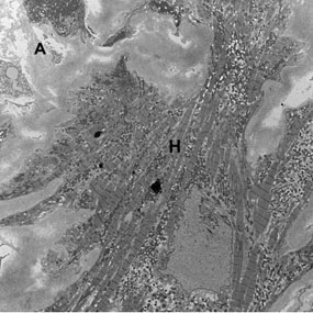
Advanced imaging called electron microscopy shows how amyloid deposits (labeled A) infiltrate normal heart muscle (labeled H).
Formation of amyloid protein deposits:
- Abnormal proteins: In many cases, amyloid protein deposits form because the protein deposited is abnormal. The protein's abnormal structure makes it more likely to form into fibrils and accumulate in body tissues.
- High quantities: In other cases, a normal protein is deposited in an organ because the protein is present in very high quantities. Or, it has been present for many years, slowly depositing over time.
These protein deposits also contain other substances. Medical experts are researching how these substances help form amyloid deposits.
Amyloidosis: Prognosis
The biggest factor in determining life expectancy for patients with amyloidosis is finding out how much the heart is involved. Each person with this disease is unique, with many factors affecting his or her prognosis.
However, even patients with advanced heart involvement can often benefit greatly from treatment at an experienced amyloid center. To learn more, speak with your physicians about your specific prognosis.
Amyloidosis Organ Involvement
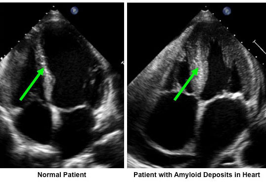
Amyloid fibrils make the heart's walls appear thicker when they accumulate in the heart. Notice how much thicker the wall is in the patient with amyloidosis (right) compared with a normal heart (left) in these ultrasound images.
Amyloid deposits may develop in one or more parts of the body, such as:
- Abdominal fat
- Bone marrow
- Gut
- Heart
- Kidneys
- Liver
- Nervous system
- Skin and soft tissue
- Tongue
Amyloidosis in the Heart
If amyloid deposits form in the heart, they affect the heart muscle's ability to relax and squeeze. Amyloid deposits can disrupt the heart's electrical system, causing the heart to beat too fast or too slow.
Common signs and symptoms of amyloid involvement in the heart include:
- Dizziness
- Fainting
- Fatigue
- Fluid retention
- Low blood pressure
- Shortness of breath
Heart complications are the most common cause of death in patients with amyloidosis. The degree to which amyloidosis affects the heart is important in determining your prognosis.
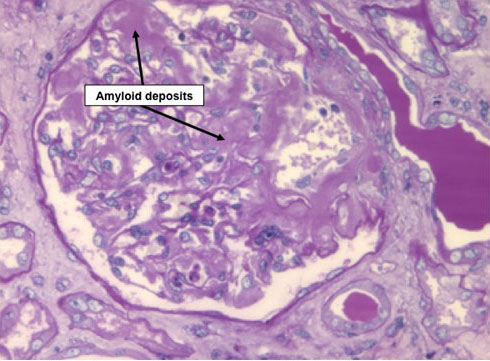
These amyloid deposits are in the kidneys. The entire filtering apparatus pictured here is flooded with amyloid deposits.
Amyloidosis in the Kidneys
Amyloid deposits commonly lead to protein loss in the urine and, in some cases, can lead to kidney failure.
Edema, or fluid retention, is a sign that your kidneys may be failing. Your ankles and/or legs may swell up as a result.
Amyloidosis in the Nerves
Amyloid deposits in the nerves lead to numbness, tingling, pain or, less commonly, weakness. In addition, amyloid deposits in the nerves may contribute to low blood pressure.
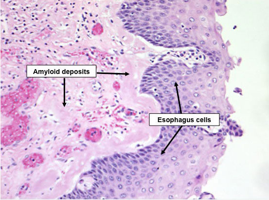
These amyloid deposits are in the esophagus of a patient with amyloidosis.
Amyloidosis in the Gastrointestinal Tract or Tongue
The gastrointestinal tract is a frequent site of amyloid deposits. Amyloid deposits can form in the tongue and cause it to enlarge, known as macroglossia.
Deposits in other areas of the gastrointestinal tract may cause:
- Bloating
- Constipation
- Diarrhea
- Difficulty swallowing
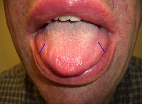
This patient has amyloid deposits in the lip and tongue. Note the scalloped appearance of the tongue in which indentations are present on both sides (arrows). The indentations occur because the enlarged tongue constantly presses against the upper teeth.
Amyloidosis in the Skin and Soft Tissue
In some people, amyloid deposits build up in the skin and soft tissue. These deposits can cause significant changes in appearance, particularly if they occur in the face.
Amyloid deposits in the wrist, along with deposits in the nerves, can lead to carpal tunnel syndrome. Amyloid deposits in the blood vessels of the skin can make them more fragile, leading to easy bruising.
Symptoms of Amyloidosis
People with this condition experience varying symptoms depending on where the amyloid deposits accumulate. Learn more about amyloidosis symptoms.
Condition Spotlight

Clinical Trials for Amyloidosis
Clinical trials are research studies that evaluate a new medical approach, device, drug, or other treatment. As a Stanford Health Care patient, you may have access to the latest, advanced clinical trials.
Open trials refer to studies currently accepting participants. Closed trials are not currently enrolling, but may open in the future.
Amyloid Center
See a Stanford specialist to learn about your treatment options. Visit our clinic to make an appointment.

Amyloidosis
Our hematologists (blood disorder specialists) offer the latest treatments for people with amyloidosis, caused by abnormal protein buildup in body organs.
Amyloidosis